Sonoanatomy for Anaesthetists
| Publisher |
Cambridge University Press |
|---|---|
| Language |
English |
| Edition |
1st |
| Format |
Publisher PDF |
| ISBN 13 |
9781139579018 |
- Best Price Guaranteed
- Best Version Available
- Free Pre‑Purchase Consultation
- Immediate Access After Purchase
$21.60
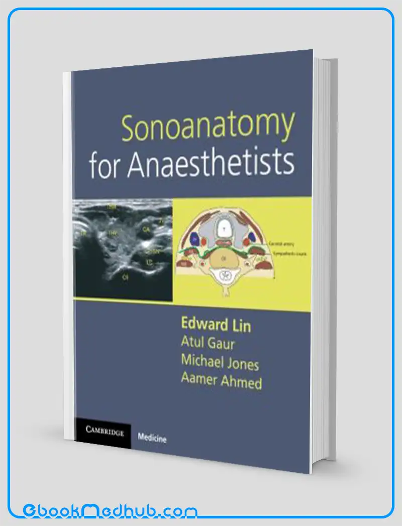
Sonoanatomy for Anaesthetists
The advancement of portable handheld ultrasound scanners has significantly enhanced the precision of clinicians in identifying nerves and blood vessels, particularly benefiting the field of anesthesia.
This comprehensive atlas of ultrasound anatomy specifically targets the primary challenges faced by individuals acquiring skills in ultrasound-guided procedures.
These challenges primarily involve determining the correct placement of the probe and understanding the anatomical structures being visualized.
For each nerve block or site for vascular access, this atlas provides a detailed depiction comprising an anatomical line illustration, a clinical photograph demonstrating the accurate positioning of the ultrasound probe, the actual ultrasound scan, and a labeled line illustration of the scan highlighting key anatomical features.
All essential anatomic regions are covered, encompassing the upper limb, lower limb, neck, thorax, and abdomen.
Succinct notes accompany each entry, indicating landmarks for scanning and offering valuable insights and recommendations on potential complications that may arise during procedures.
“Sonoanatomy for Anesthetists” emerges as a crucial reference tool for professionals in the fields of anesthesia, intensive care, and chronic pain management. Anesthetists, intensivists, and chronic pain specialists stand to benefit significantly from the wealth of information and guidance provided in this resource.
Sonoanatomy for Anaesthetists
Key Features
“Sonoanatomy for Anaesthetists” encompasses several key features:
Functionality as a Practical Atlas: The book functions as a practical atlas of ultrasound anatomy specifically tailored to aid clinicians in accurately locating nerves and blood vessels while performing ultrasound-guided procedures.
The detailed visual representations and explanations serve as a valuable tool in enhancing procedural accuracy and efficacy.
In-Depth Coverage: The comprehensive nature of “Sonoanatomy for Anaesthetists” ensures that it covers all pertinent anatomic regions, such as the upper limb, lower limb, neck, thorax, and abdomen.
This wide-ranging coverage expands the applicability of the book to various scenarios encountered in anaesthesia practice, thus providing a thorough resource for practitioners.
Guided Visual Assistance: Each section dedicated to a nerve block or vascular access site is supplemented with an anatomical line illustration, a clinical photograph illustrating the correct positioning of the ultrasound probe, the corresponding ultrasound scan, and a labelled line illustration highlighting crucial anatomical landmarks.
This visual guidance aids practitioners in accurately identifying and navigating anatomical structures during procedures.
Concise Information: “Sonoanatomy for Anaesthetists” includes succinct notes accompanying each entry, offering valuable insights on scan landmarks, tips, and advice on potential complications.
These concise notes aim to enrich the reader’s comprehension and practical skills, thereby enhancing their ability to perform ultrasound-guided procedures effectively.
Vital Educational Tool: Serving as an indispensable resource, “Sonoanatomy for Anaesthetists” caters to anaesthetists, intensivists, and chronic pain specialists, facilitating their mastery of ultrasound-guided techniques.
By equipping practitioners with the knowledge and skills needed to conduct safe and efficient procedures, this book plays a pivotal role in enhancing clinical practice and patient outcomes.

Sonoanatomy for Anaesthetists
Summary
“Sonoanatomy for Anaesthetists” is a valuable and practical guide for healthcare professionals aiming to enhance their skills in performing ultrasound-guided procedures.
The introduction of portable handheld ultrasound devices has made the precise identification of nerves and blood vessels crucial in the field of anesthesia.
The book covers important anatomical regions such as the upper limb, lower limb, neck, thorax, and abdomen. Each section dedicated to a nerve block or vascular access point is meticulously detailed with anatomical line drawings, clinical images showing the correct placement of the ultrasound probe, corresponding ultrasound scans, and labeled illustrations pointing out key anatomical landmarks.
In addition to the comprehensive visuals, “Sonoanatomy for Anaesthetists” also provides concise notes for each topic, offering advice on where to focus during the scan and valuable insights on potential complications.
This resource is essential for anaesthetists, intensivists, and chronic pain specialists looking to improve their ultrasound-guided techniques and ensure the delivery of safe and efficient patient care.

Sonoanatomy for Anaesthetists
This website offers ( Sonoanatomy for Anaesthetists ) with just a few clicks.
The website strives to provide you with simple access to the medical field as well as readily available information that you can download.
You can download all of the books at a reasonable price and get the most recent scientific data in the world of medicine anytime you want at ebookmedhub.com.
Other Products :
Rubins Pathology Clinicopathologic Foundations of Medicine 7th Edition (ORIGINAL PDF from Publisher)
Robbins and Cotran Pathology Flash Cards (ORIGINAL PDF from Publisher)
Radiographic Pathology Workbook 2nd Edition (ORIGINAL PDF from Publisher)





Reviews
There are no reviews yet.