Osborns Brain Imaging Pathology and Anatomy (High Quality CHM)
| Publisher |
Lippincott Williams & Wilkins |
|---|---|
| Language |
English |
| Edition |
1st |
| Format |
HQ CHM |
| ISBN 10 |
1931884218 |
| ISBN 13 |
978-1931884211 |
- Best Price Guaranteed
- Best Version Available
- Free Pre‑Purchase Consultation
- Immediate Access After Purchase
$28.00
Osborns Brain Imaging Pathology and Anatomy (High Quality CHM)
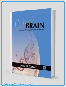
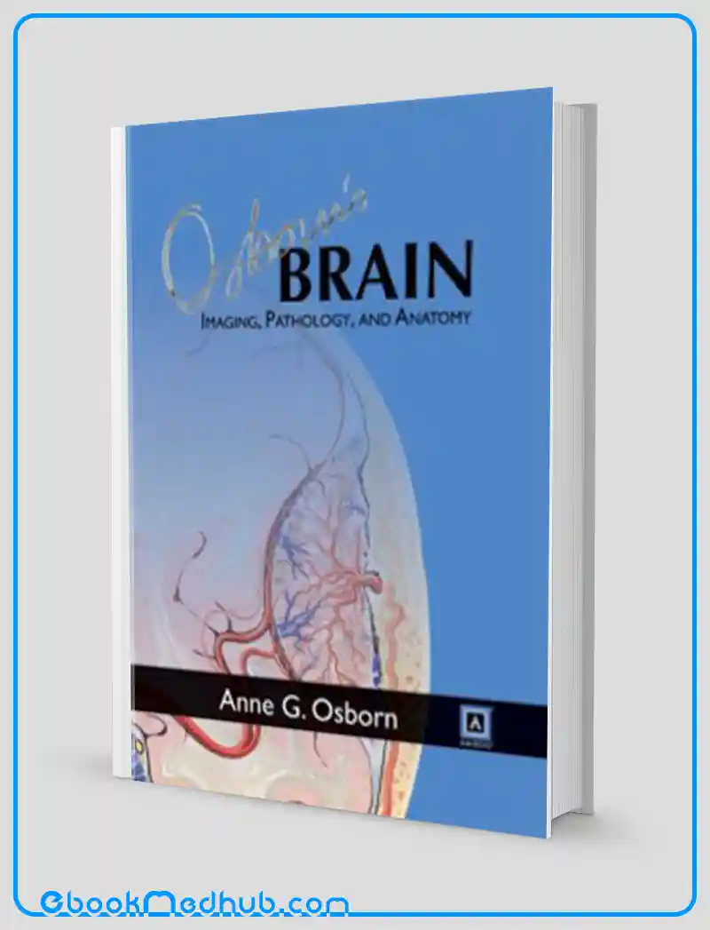
Anne G. Osborn MD FACR presents Osborn’s Brain: Imaging, Pathology, and Anatomy, which has been eagerly awaited as the successor to her renowned 1993 book Diagnostic Neuroradiology (also known as “The Red Book”), a highly popular text in the field of neuroradiology. The previous book became a bestseller over time, capturing the attention of many readers.
This 1,200-page volume by Anne Osborn is highly anticipated due to her unique approach in making intricate subjects visually appealing and easily comprehensible.
By combining essential brain imaging knowledge with remarkable pathology examples, relevant anatomy details, and the latest brain imaging techniques, Osborn offers a comprehensive resource for readers.
Osborn’s Brain is structured to support curriculum-based learning, beginning with crucial topics like trauma to facilitate practical learning for residents or practicing radiologists. Through this organization, readers can grasp the thought processes involved in making diagnoses, understanding different types of diagnoses, and recognizing the diverse brain pathologies.
Osborns Brain Imaging Pathology and Anatomy (High Quality CHM)
While guiding readers through the realm of brain imaging, Osborn introduces novel concepts and diagnoses that will captivate even the most experienced neuroradiologists.
The book’s format differs significantly from Osborn’s recent works, moving away from bulleted text to detailed prose paragraphs aimed at providing a thorough learning experience. Additionally, summary boxes are included to emphasize key points in each section, enhancing usability and functionality.
The book’s content is enriched with over 3,300 high-resolution radiologic images and medical illustrations, all meticulously annotated to highlight clinically significant features.
Osborn’s Brain is not only informative but also visually engaging, offering clear and concise information in an easily digestible format. This remarkable volume delivers essential knowledge for radiologists and clinical neuroscientists, taking them on an intriguing journey through the complexities of Osborn’s brain.

Osborns Brain Imaging Pathology and Anatomy (High Quality CHM)
Key Features
The book “Osborns Brain Imaging Pathology and Anatomy (High Quality CHM)” encompasses key features that distinguish it from other works in the field:
As the eagerly anticipated successor to the highly esteemed “Diagnostic Neuroradiology” by Anne G. Osborn, a text affectionately known as “The Red Book,” this volume upholds the rich legacy of one of the most widely acclaimed neuroradiology texts of all time.
The enduring success of Osborns Brain Imaging Pathology and Anatomy can be attributed to the extensive expertise of Anne Osborn herself, who presents intricate subjects in a visually captivating and easily comprehensible manner, ensuring that readers can grasp even the most complex concepts with relative ease.
One of the standout features of Osborns Brain Imaging Pathology and Anatomy is its comprehensive nature, which seamlessly integrates fundamental brain imaging principles, illustrative pathology examples, pertinent anatomical insights, and the latest advancements in brain imaging techniques, providing readers with a vast array of knowledge that is essential for a well-rounded understanding of the subject matter.
Moreover, the book is structured to facilitate curriculum-based learning, commencing with critical topics such as trauma, a deliberate approach that assists readers in acquiring knowledge in a logical sequence that is particularly beneficial for residents and practicing radiologists alike.
In addition to imparting foundational knowledge, Osborn emphasizes the importance of cultivating a profound insight into diagnostic thinking, different types of diagnoses, and the diverse spectrum of brain pathologies encountered in clinical practice.
Unlike conventional textbooks that rely on bullet points, Osborns Brain Imaging Pathology and Anatomy adopts a prose format consisting of detailed paragraphs, thereby enhancing its value as a comprehensive educational tool that promotes in-depth learning and understanding.
Each section of the book incorporates summary boxes that succinctly encapsulate the most essential points, thereby enhancing the book’s usability and facilitating better retention of key information.
Furthermore, the visual richness of Osborns Brain Imaging Pathology and Anatomy is underscored by its inclusion of over 3,300 high-resolution radiologic images and medical illustrations, providing readers with a visually immersive and informative experience that complements the textual content.
Moreover, all images and illustrations within the book are meticulously annotated to draw attention to clinically significant features, thereby aiding readers in better comprehension of the material presented.
Designed to be user-friendly, visually stimulating, and packed with concise, clear information, Osborns Brain Imaging Pathology and Anatomy ensures that readers can engage with the content effortlessly and derive maximum benefit from their reading experience.
Lastly, Osborns Brain Imaging Pathology and Anatomy offers readers a fascinating journey into the vast knowledge and expertise of Anne Osborn, providing a captivating exploration of the intricacies of brain imaging and pathology that caters to the specific needs of radiologists and clinical neuroscientists seeking to deepen their understanding of the subject matter.

Osborns Brain Imaging Pathology and Anatomy (High Quality CHM)
This website offers ( Osborns Brain Imaging Pathology and Anatomy (High Quality CHM) ) with just a few clicks.
The website strives to provide you with simple access to the medical field as well as readily available information that you can download.
You can download all of the books at a reasonable price and get the most recent scientific data in the world of medicine anytime you want at ebookmedhub.com.
Other Products :
Anatomy Essentials For Dummies (Original PDF from Publisher)
Anatomy An Essential Textbook An Illustrated Review (Original PDF from Publisher)
Anatomic Basis of Neurologic Diagnosis (Original PDF from Publisher)



Reviews
There are no reviews yet.