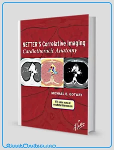
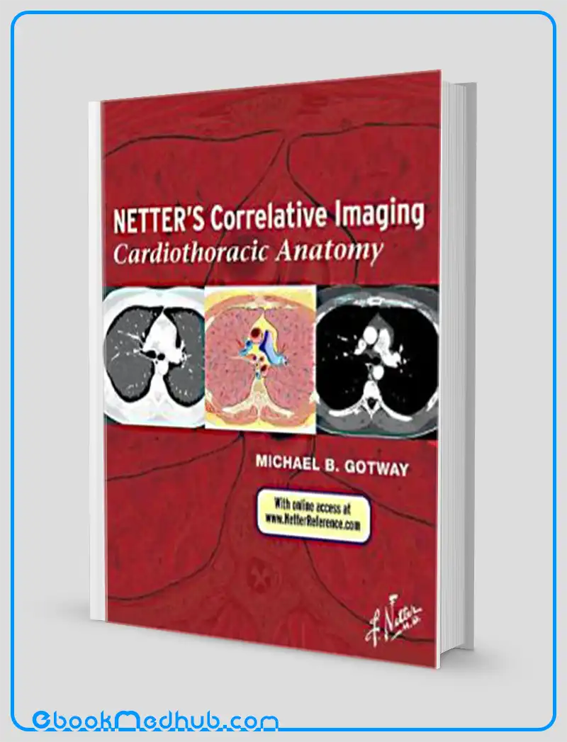
Netters Correlative Imaging Cardiothoracic Anatomy (ORIGINAL PDF from Publisher)
| Publisher |
Elsevier |
|---|---|
| Language |
English |
| Edition |
1st |
| Format |
Publisher PDF |
| ISBN 13 |
9781437704402 |
- Best Price Guaranteed
- Best Version Available
- Free Pre‑Purchase Consultation
- Immediate Access After Purchase
$29.60
Categories: AnatomyCardiologyHot BooksRespiratory Medicine
Netters Correlative Imaging Cardiothoracic Anatomy (ORIGINAL PDF from Publisher)
Cardiothoracic Anatomy, the third volume in the newly introduced Netter’s Correlative Imaging series, offers exceptional visual guidance on thoracic, chest wall, lung, and heart anatomy.
Dr. Michael Gotway skillfully presents Netter’s exquisite and educational paintings and illustrated cross sections following the distinctive Netter style, placed alongside high-resolution patient images obtained from breath-hold cardiac MR, multislice thoracic CT, and CT coronary angiography techniques.
This juxtaposition aids in a comprehensive visualization of the anatomy, section by section, allowing for a deeper understanding of the intricate structures involved.
The detailed coverage provided, along with succinct descriptive text for quick reference, offers immediate access to essential information, making it an invaluable resource for today’s imaging specialists who are constantly pressed for time.
Examine thoracic, chest wall, lung, and heart anatomy through the lens of breath-hold cardiac MR, multislice thoracic CT, and CT coronary angiography, with each image thoughtfully paired with a meticulously crafted illustration designed in the educational yet visually appealing Netter style.
Easily identify anatomical landmarks with the help of comprehensive labeling and succinct text that directs attention to key points relevant to the paired illustration and image.
Access the correlated images conveniently online at www.NetterReference.com, allowing for a seamless transition between the detailed illustrations and the corresponding patient images.
This online feature enhances the learning experience by providing a dynamic platform for further exploration and reference beyond the physical pages of the atlas, catering to the needs of modern imaging professionals seeking a comprehensive and accessible anatomical resource.
Netters Correlative Imaging Cardiothoracic Anatomy (ORIGINAL PDF from Publisher)
Key Features
Key features of “Netters Correlative Imaging Cardiothoracic Anatomy” encompass a wide range of elements. The book offers a thorough exploration of thoracic, chest wall, lung, and heart anatomy, establishing itself as a comprehensive point of reference for professionals in the field of medical imaging.
It combines Netter’s renowned illustrations with high-resolution patient images derived from cutting-edge imaging technologies like breath-hold cardiac MR, multislice thoracic CT, and CT coronary angiography.
Each illustration in the book is meticulously labeled and accompanied by succinct text that emphasizes crucial anatomical landmarks, facilitating an easy and efficient process of identification and comprehension for the readers.
Furthermore, the inclusion of online resources accessible at www.NetterReference.com serves to enhance the book’s usability by providing readers with additional correlated images, thereby enriching their visual learning experience.
Specifically tailored to cater to the demands of busy imaging specialists, “Netters Correlative Imaging Cardiothoracic Anatomy” offers a swift and convenient route to pertinent anatomical information, making it an ideal companion for professionals navigating the complexities of medical imaging on a daily basis.

Netters Correlative Imaging Cardiothoracic Anatomy (ORIGINAL PDF from Publisher)
Summary
“Netters Correlative Imaging Cardiothoracic Anatomy” introduces an impressive combination of Dr. Michael Gotway’s cardiothoracic radiology expertise and the renowned Netter style of anatomical illustration.
This resource offers a distinctive visual aid for comprehending the complex anatomy of the thoracic area, covering structures like the chest wall, lungs, and heart in detail.
The book effectively merges high-resolution patient images obtained from advanced imaging techniques such as breath-hold cardiac MR and multislice thoracic CT with meticulously crafted Netter illustrations, providing a superior learning tool for medical professionals aiming to strengthen their understanding and interpretation of cardiothoracic anatomy.
Through detailed labeling and concise explanatory content, Netters Correlative Imaging Cardiothoracic Anatomy facilitates swift and precise identification of anatomical landmarks crucial for accurate diagnosis and treatment planning.
Moreover, the supplementary online access to correlated images elevates the book’s usefulness, establishing it as an essential reference for busy imaging specialists and healthcare practitioners working within the radiology field.
This comprehensive resource not only serves as a valuable educational tool but also as a practical aid in the day-to-day practice of healthcare professionals involved in cardiothoracic imaging.

Netters Correlative Imaging Cardiothoracic Anatomy (ORIGINAL PDF from Publisher)
This website offers ( Netters Correlative Imaging Cardiothoracic Anatomy (ORIGINAL PDF from Publisher) ) with just a few clicks.
The website strives to provide you with simple access to the medical field as well as readily available information that you can download.
You can download all of the books at a reasonable price and get the most recent scientific data in the world of medicine anytime you want at ebookmedhub.com.
Other Products :
Nevos Tumores Neviformes y Sindromes Nevicos (Spanish Edition) (High Quality Image PDF)
Pain A textbook for health professionals 3rd edition (Original PDF from Publisher)
Physician Assistant Board Review 4th edition (Original PDF from Publisher)

















Reviews
There are no reviews yet.