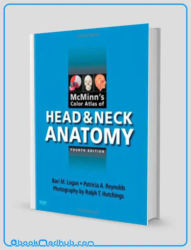
McMinns Color Atlas of Head and Neck Anatomy 4e
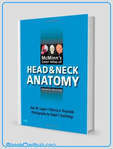
McMinn’s Color Atlas of Head and Neck Anatomy is an exceptional large format atlas showcasing detailed dissections, osteology, and radiographic as well as surface anatomy images of the human head and neck.
Authored by Bari M. Logan, Patricia Reynolds, and Ralph T. Hutchings, this atlas serves as a comprehensive “road map” reference for the anatomical structures within the head and neck region, making it an invaluable resource for both study purposes and exam revision.
The latest edition of the atlas includes new dissections and double-page spreads that offer in-depth depictions of specific areas, enhancing the overall content coverage. Complex dissections are supported by explanatory artwork, while all dissections come with accompanying notes and commentaries, enriching the reader’s understanding of the anatomical structures presented.
McMinns Color Atlas of Head and Neck Anatomy 4e
Furthermore, reference lists and dental anesthesia information are conveniently located in the appendices at the end of the book, providing additional resources and clinical insights. In essence, this atlas stands out as a visually stunning and comprehensive resource for the entire head and neck region.
“It is challenging to identify any shortcomings in a meticulously crafted book that remains highly relevant in its comprehensiveness. Merely having a copy in the library for occasional reference is insufficient – every individual should possess their personal copy!”
The European Journal of Orthodontics, in its review from July 2010, highlighted the following key features of the atlas: it offers life-sized images of dissections and osteology that correspond to real-life observations in laboratory settings or clinical practice.
Additionally, the inclusion of radiographic and surface anatomy pictures ensures the clinical relevance of the content. Detailed notes and commentaries are provided for each dissection, along with orientational and explanatory artwork for complex structures, aiding in a complete comprehension of positioning and application.
Moreover, the incorporation of reference lists and dental anesthesia information in the appendices at the end of the book serves as a valuable resource for further study and clinical relevance. The atlas also introduces 12 new “in-fill” dissections focusing on individual glands, establishing itself as the definitive reference for head and neck anatomy.
Furthermore, new double-page spreads cover topics such as the vascularity of the brain, eruption and growth of deciduous teeth, sutural bones, and the skull, offering detailed depictions of these crucial areas. Clinical photographs reflecting current practices have been added to keep readers abreast of the latest developments in the field.

McMinns Color Atlas of Head and Neck Anatomy 4e
Key Features
The “McMinns Color Atlas of Head and Neck Anatomy 4e” is characterized by several key features:
One of the primary aspects of this atlas is its comprehensive coverage of human head and neck anatomy, catering to the needs of both students and professionals in the field. The extensive coverage makes it an invaluable resource for individuals seeking a deep understanding of this intricate area of the human body.
Moreover, the large format in which McMinns Color Atlas of Head and Neck Anatomy 4e is presented allows for detailed, clear, and visually appealing illustrations and images. This facilitates a more in-depth exploration and comprehension of the anatomical structures depicted in the atlas.
Furthermore, the inclusion of radiography and surface anatomy images adds a layer of clinical relevance to the content, making it directly applicable to real-world practice scenarios. This ensures that learners can connect theoretical knowledge with practical applications in various medical settings.
Additionally, the atlas features life-sized images of dissections and osteology, corresponding closely with what is typically observed in laboratory or clinical practice settings. This aspect provides learners with a realistic and accurate representation of anatomical structures, aiding in their learning and comprehension process.
Complex dissections within the atlas are further elucidated by explanatory artwork, notes, and commentaries, which serve to enhance the understanding of anatomical positions and their practical applications. This additional context helps learners grasp the intricacies of anatomical structures more effectively.
Moreover, the inclusion of appendices containing reference lists and dental anesthesia information at the back of the atlas offers readers additional resources and clinical material to supplement their learning experience. These supplementary materials provide further depth and context to the information presented in the main body of the atlas.
The fourth edition of McMinns Color Atlas of Head and Neck Anatomy introduces new dissections, double-page spreads on key areas, and updated clinical photographs to align with current practices in the field. This ensures that learners have access to the most up-to-date information and visuals related to head and neck anatomy.
Lastly, the atlas offers 12 new “in-fill” dissections of single glands, serving as a definitive reference for individuals studying head and neck anatomy. This detailed and in-depth content caters to the needs of learners looking to deepen their knowledge and understanding of specific anatomical structures within the head and neck region.

McMinns Color Atlas of Head and Neck Anatomy 4e
This website offers ( McMinns Color Atlas of Head and Neck Anatomy 4e ) with just a few clicks.
The website strives to provide you with simple access to the medical field as well as readily available information that you can download.
You can download all of the books at a reasonable price and get the most recent scientific data in the world of medicine anytime you want at ebookmedhub.com.
Other Products :
Anatomic Basis of Neurologic Diagnosis (Original PDF from Publisher)
Anatomy An Essential Textbook An Illustrated Review (Original PDF from Publisher)
Anatomy Essentials For Dummies (Original PDF from Publisher)

















Reviews
There are no reviews yet.